“Saving and Loading” Nerve Signals

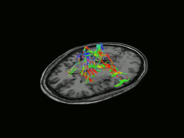
Electrical stimulation of peripheral nerves can treat patients without necessitating pharmaceutical drugs. Stimulation of the vagus nerve, for instance, treats epilepsy, depression, arthritis, IBS, and more. However, nerve stimulation can lead to side effects when done improperly—that’s why we are working to optimize “Spatially Selective” stimulation, which delivers electricity to only the locations in a nerve which control desired effects. We use a multi-contact cuff electrode for stimulation and recording.

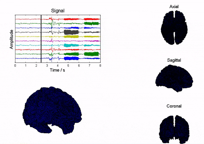
We have developed two algorithms which optimize spatially selective stimulation. First, we determine where the electrical current needs to be delivered, using a combination of imaging tools and custom source localization algorithms. Secondly, we determine the correct amount of current to apply to each electrode to precisely activate the target locations on the nerve. Our in-Silico and physical model findings demonstrate that we can use recording and stimulation together, to “save” a neural signal, and “load” that signal later

“Brain Copying”

In the field of neuroscience, we all dream of direct brain-to-brain communication. Our lab is paving the way towards making that dream into a reality.

By combining brain recording tools on one individual with brain stimulation methodologies on another individual, we are working towards “copying” a signal from one brain into another. By recording activity in the first subject’s brain, we can find the origin of each signal—then use stimulation to apply electrical activity to the same region of the second subject’s brain. This entire process can be performed non-invasively.

Our group has demonstrated the precision of our stimulation and recording techniques in-Silico, and we are working on the development of a stimulator that can be used to test out “Brain Copying” on animal models.

Brain Copying Fundamentals:
Brain Copying requires cutting-edge recording and stimulation tools. Our lab has developed both (images from our publications https://doi.org/10.3389/fnins.2016.00543, and https://doi.org/10.3390/a15050169)


Brain Dynamics and Source Localization

Brain imaging techniques can help us explore the function of human brain (i.e. memory, behavior, etc.) and help diagnosis and treatment of brain disorders (i.e. seizure, depression, etc.). Imaging tool with high temporal and spatial resolution is highly desirable. Currently, fMRI is very popular since it offers high spatial resolution (~3mm); however, its temporal resolution is limited (~1s) and is impossible to be used in portable applications. EEG has excellent temporal resolution (~1ms), portable mobility that allowed to be used in daily life. Our goal here is to improve the spatial resolution of EEG to be as good as that of fMRI.

source-localization-2-gif-1

We study the brain dynamics and source localization based on realistic head model (NFT toolbox). On one hand, we are studying the influence of various factors (i.e. noise, electrode number, head model, inverse solution, etc.) on the accuracy of inverse imaging; on the other hand, we are developing more accurate inverse imaging algorithms.


Collaborators


  1. Prof. Scott Makeig, SCCN, UCSD
  2. Dr. Yue-Loong Hsin, Chung Shan Medical University, Taiwan