Stimulation Artifact Cancellation

2016-07-01_15_04_54_02sec-pageLive Stimulation Artifact Cancellation


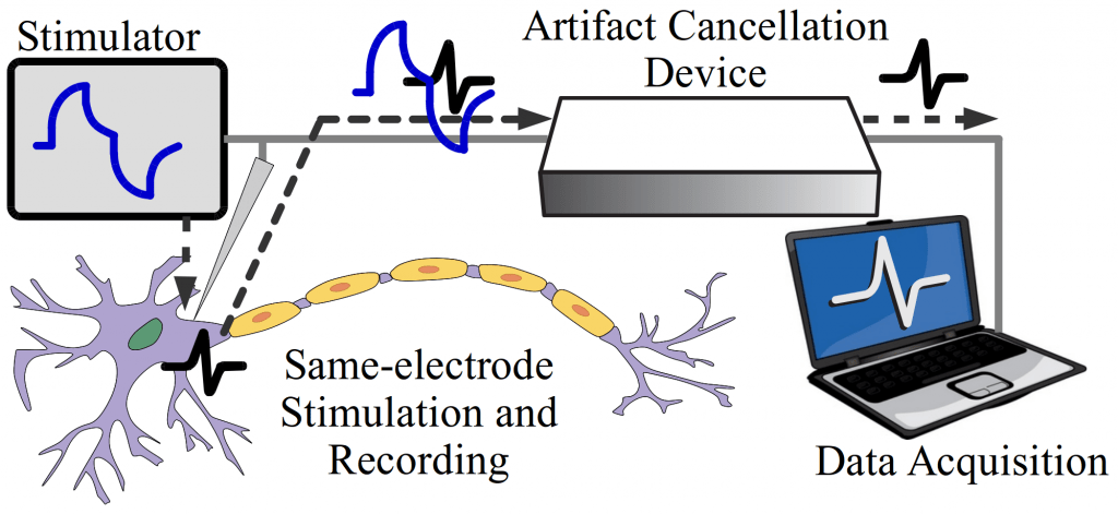
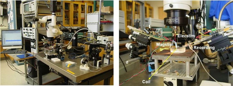
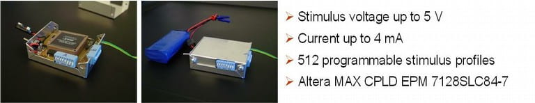
Recovering neural responses from electrode recordings is fundamental for understanding the dynamics of neural networks. This effort is often obscured by stimulus artifacts in the recordings, which result from stimuli injected into the electrode-tissue interface. Stimulus artifacts, which can be orders of magnitude larger than the neural responses of interest, can mask short-latency evoked responses. Furthermore, simultaneous neural stimulation and recording on the same electrode generates artifacts with larger amplitudes compared to a separate electrode setup, which inevitably overwhelm the amplifier operation and cause unrecoverable neural signal loss. This paper proposes an end-to-end system combining hardware and software techniques for actively cancelling stimulus artifacts, avoiding amplifier saturation, and recovering neural responses during current-controlled in-vivo neural stimulation and recording. The proposed system is tested in-vitro under various stimulation settings by stimulating and recording on the same electrode with a superimposed pre-recorded neural signal. Experimental results show that neural responses can be recovered with minimal distortion even during stimulus artifacts that are several orders greater in magnitude.


Artifact Cancellation fig1_systemOverview copy
System-Level Overview


Publication

“A Hybrid Hardware and Software Approach for Cancelling Stimulus Artifacts During Same-electrode Neural Stimulation and Recording,” Stanislav Culaclii, Brian Kim, Yi-Kai Lo, and Wentai Liu, accepted by EMBC 2016.

Cross Frequency Coupling (CFC)


CFC-3


Cross Frequency Coupling (CFC) is the interaction between brain oscillations of different frequencies, and the coupling phenomenon has been observed in the brain of rodent and human. Phase-amplitude coupling (PAC) is a type of CFC, which describes the dependence between the phase of a low-frequency component and the amplitude of a high-frequency component of electrical brain activities. It has been claimed that the modulation of low frequency phase on high frequency amplitude plays a functional role in cognition and information processing, such as learning and memory. The change of PAC patterns has been associated with various neurological disorders, e.g., epilepsy and  schizophrenia.

Five seizure stages classified by firing patterns (on the left). PAC pattern comparisons between the conventional method (middle) and HHT method (right) of two patients.

Non-stationary and nonlinear techniques for seizure detection algorithms

Epilepsy is one of the most common neurological diseases, affecting over 3 million people in U.S. and 50 million (~1%) people worldwide. An automated and accurate seizure detection method can be very helpful. Currently, most people use stationary and linear methods (i.e. Fast Fourier Transform (FFT), etc.) to analyze the signal. However, EEG signal is non-stationary and nonlinear in nature, thus these methods will introduce inaccuracy. Hence, we are developing non-stationary and nonlinear algorithms to improve the accuracy of seizure detection.
Corr3


Collaborators


  1. Dr. Yue-Loong Hsin, Chung Shan Medical University, Taiwan

Electrode-based Bio-imaging

Our goal is to develop an Electrode-based Bio-imaging System that will be fMRI-competitive. Currently, fMRI is very popular since it offers high spatial resolution (~3mm); however, its temporal resolution is limited (~1s) and is impossible to be used in portable applications. EEG has excellent temporal resolution (~1ms), portable mobility that allowed to be used in daily life. Here, we devote to improving the spatial resolution of EEG in the aspects of both hardware and software.
 
1. Hardware
We developed a Focused Electrode that improves spatial resolution, recording SNR and reduce crosstalk and correlation with adjacent electrodes. The Focused Electrode is adaptive to the geometry parameters of electrode array including electrode size, pitch and source depth, such as EEG or ECoG. Simulation results show that the Focused Electrode increases the number of electrodes up to 7x in EEG and 30x in ECoG without overlapping information if the array covers half of head.

300px-Model
2. Software
We study the inverse imaging using realistic head model based on MR image (NFT toolbox). On one hand, we are studying the influence of various factors (i.e. noise, electrode number, head model, inverse solution, etc.) on inverse imaging; on the other hand, we are developing more accurate inverse imaging algorithms.


source-localization-gif-2


Collaborators


  1. Prof. Scott Makeig, SCCN, UCSD
  2. Dr. Yue-Loong Hsin, Chung Shan Medical University, Taiwan

Cardiac Myocyte Stem Cell Differentiation

We collaborate with Dr. Peyman Benharash, Department of Surgery, UCLA in stem cell differentiation with the help of electrical stimulation. The following is a demonstrational video of Cardiac Myocyte Cells. The sorting result shows that couple cell groups are beating separately. The cells is anticipated to be eventually grouped and beating together and form a patch for cardiac disease repair.

Myocyte_sorting

Brain Dynamics and Source Localization

Brain imaging techniques can help us explore the function of human brain (i.e. memory, behavior, etc.) and help diagnosis and treatment of brain disorders (i.e. seizure, depression, etc.). Imaging tool with high temporal and spatial resolution is highly desirable. Currently, fMRI is very popular since it offers high spatial resolution (~3mm); however, its temporal resolution is limited (~1s) and is impossible to be used in portable applications. EEG has excellent temporal resolution (~1ms), portable mobility that allowed to be used in daily life. Our goal here is to improve the spatial resolution of EEG to be as good as that of fMRI.

source-localization-2-gif-1

We study the brain dynamics and source localization based on realistic head model (NFT toolbox). On one hand, we are studying the influence of various factors (i.e. noise, electrode number, head model, inverse solution, etc.) on the accuracy of inverse imaging; on the other hand, we are developing more accurate inverse imaging algorithms.


Collaborators


  1. Prof. Scott Makeig, SCCN, UCSD
  2. Dr. Yue-Loong Hsin, Chung Shan Medical University, Taiwan

Neural Signal Processing and Telemetry

Recording from a large number of neurons produces vast quantities of data that is highly difficult to extract and interpret due to noise and aggregation of multiple neural signals in a single recording site. In addition to conventional techniques like spike sorting, component analysis, de-noising detection, we are focusing on the cellular and molecular levels to understand the fundamentals of neural signals and behavior of large group of neurons. We found that the traditional deterministic channel models are insufficient for the description of the activities of real neurons. We are currently developing the stochastic kinetic modeling of sodium channel and its validation with the measurement of gate current due to transmembrane protein movements in the order of several picoamps with the bandwidth of several hundred of MHz.


Selected Publications


  1. “A 12-channel 6mW Wireless Neural Recording IC with On-the-fly Spike Sorting and UWB Transmitter,” M. Chae, W. Liu, Z. Yang, T. Chen, J. Kim, M. Sivaprakasam, and M. R. Yuce, To appear in International Solid-State Circuits Conference, February 2008.
  2. “An Integrated Multi-Channel Neural Recording System,” M. Chae, W. Liu, and M. Sivaprakasam, BMES Annual Fall Meeting, September 2007.


Collaborators


  1. Huntington Medical Research Institutes
  2. Arizona State University

Non-Invasive Functional Magnetic Stimulation

Neural stimulation is commonly accomplished by a voltage or current pulse through a microelectrode. Ideally, a method would exist which inherently had zero net charge transfer, required only simple driver circuitry and was completely isolated from the tissue to reduce circuit failure due to corrosion and fouling by protein deposition. Magnetic stimulation achieves these goals. The presence of scar tissue or deposited proteins is irrelevant because the magnetic permeability of tissue is near that of free space. Excitation arises from the magnetic field which generates a current across the membrane of the cell which changes the resting potential of the neuron and triggers an action potential (the fundamental signal generation mechanism neurons employ). We are currently studying the fundamental mechanisms of magnetic stimulation, developing models and verifying through in vitro experiments.
Magstim

Magrecording


Selected Publications


  1. “Functional Magnetic Stimulation for Epiretinal Prosthesis”, E. Basham, W. Liu, and M. Sivaprakasam, Abstract B254, Association of Research in Vision and Ophthalmology Annual Meeting, May 2005.


Collaborators


  1. Santa Clara University

Microstimulation Platform for Neural Systems

Functional Electrical Stimulation of biological tissue has a wide range of applications ranging from pain relief to neural prostheses. Flexibility, small size and low power operation, safety are key requirements in microstimulation systems. Custom integrated circuits for microstimulation face the challenge of having to support relatively high stimulation voltages for the current CMOS technology, while still needing maintain low power operation and achieve a high degree of miniaturization. In addition, the experimental nature of the evolving microstimulation applications demands a high degree of flexibility and versatility. We are working on several microstimulation applications through our collaborators that have a varying degree of requirements.
Stimchip

Wearablestim

Selected Publications


  1. “Challenges for Integrated Circuits in Implantable Devices,” W. Liu and M. Sivaprakasam, Volume: 20, Future Fab International, January 2006.
  2. “Microelectronics Design for Implantable Wireless Biomimetic Microelectronic Systems,” W. Liu, M. Sivaprakasam, G. Wang, M. Zhou, J. Granacki, J. LaCoss, and J. Wills, Volume: 24, Pages: 66 – 74, IEEE Engineering in Medicine and Biology Magazine, September 2005.
  3. “A Variable Range Bi-Phasic Current Stimulus Driver Circuitry for an Implantable Retinal Prosthetic Device,” M. Sivaprakasam, W. Liu, M. S. Humayun, and J. D. Weiland, Volume: 41, Pages: 763 – 771, IEEE Journal of Solid State Circuits, March 2005.

Collaborators


  1. University of Southern California
  2. Long Beach Veteran Affairs
  3. Stanford University
  4. Huntington Medical Research Institutes

Low Power Integrated Neural Recording System

Advances in micro electrode arrays (MEA) have enabled neuroscientists and researchers in biomedical engineering to take advantage of a large number of channels, and this has made it possible to pursue a variety of neuroprosthetic applications such as treating spinal cord injuries, deep brain stimulation to treat Parkinson’s disease. This also requires neural recording electronics that are small enough to be integrated close to the MEA and consume less power so that they can be safely placed close to the tissue. Though several efforts have been made by the research community to minimize the power and area of each individual circuit block, almost no attention was paid to find the trade-offs among those circuit blocks to achieve an optimal design. We are currently developing optimal design methodologies for integrated neural recording systems that allow the optimal design for a given set of specifications and constraints. These are verified by custom chip fabrication and verification. Wireless telemetry is essential for recording neural signals from the subject in its natural environment. We are currently developing wireless transceivers for short range telemetry for neural signals. Power and area are the key parameters for the transceivers to be implantable. We are investigating different architectures and the associated tradeoffs between computation and communication.
Neuralrecording2


Selected Publications


  1. “A 12-channel 6mW Wireless Neural Recording IC with On-the-fly Spike Sorting and UWB Transmitter,” M. Chae, W. Liu, Z. Yang, T. Chen, J. Kim, M. Sivaprakasam, and M. R. Yuce, International Solid-State Circuits Conference, February 2008.
  2. “An Integrated Multi-Channel Neural Recording System,” M. Chae, W. Liu, and M. Sivaprakasam, BMES Annual Fall Meeting, September 2007.
  3. “A Wideband Telemetry Unit for Multi-Channel Neural Recording,” M. R. Yuce, W. Liu, M. Chae, and J. Kim, IEEE International Conference on Ultra-Wideband, September 2007.


Collaborators


  1. Huntington Medical Research Institutes
  2. Arizona State University