DEMONSTRATION OF CLOSED-LOOP STIMULATION PLATFORM WITH HARDWARE ACCELERATED ARTIFACT REMOVAL

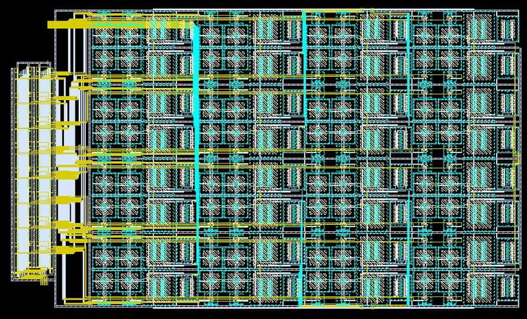
The lab has developed and validated a closed-loop stimulation platform capable of updating the stimulation montage at intervals as brief as 10 milliseconds, accompanied by a latency of only 20 milliseconds. However, the frequent updates to the montage generate biomimetic and varying electrical artifacts, thereby obscuring the neural response. Our research team has designed a proprietary integrated chip that leverages the spatial correlation inherent in the artifacts to facilitate hardware-accelerated removal. It enables uninterrupted, real-time monitoring of neural responses during active stimulation.

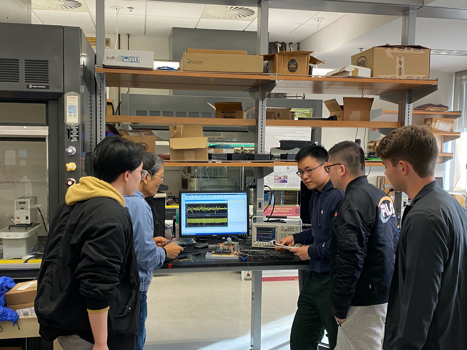
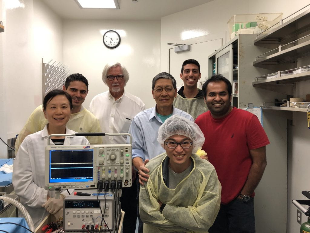
The lab member is validating the recording system’s capability to record emulated neural signals concurrently with biomimetic stimulation, specifically dynamic stimulation. The upper trace displays on the screen represents the raw input data, which includes stimulation artifacts and an oscillatory signal representing the underlying neural response. The bottom trace displays the neural response extracted by the proprietary recording system.

Demonstration of Closed-loop Operation with Our Implantable Device


The blue and pink traces on the oscilloscope represents two of the many channels that are generating stimulation pulses, initially at 30Hz and 60Hz at different phase delays respectively. The green trace represents ADC recording that is wirelessly transmitted to a laptop running the closed-loop controller in real-time. Toggling the device’s analog recording sensor, the closed-loop controller on the laptop detects the change and modulates the stimulation protocols, communicating this back to the SoC chip wirelessly. The last portion of the video shows the new stimulation patterns of the blue and pink traces, at 30Hz and 20Hz, and phase delays respectively. In summary, we have demonstrated our system’s capability to simultaneously perform real-time recording and stimulation. Accordingly, it is feasible to implement closed-loop operations using our implantable system. A sophisticated controller is under development.

Click to view the Demo

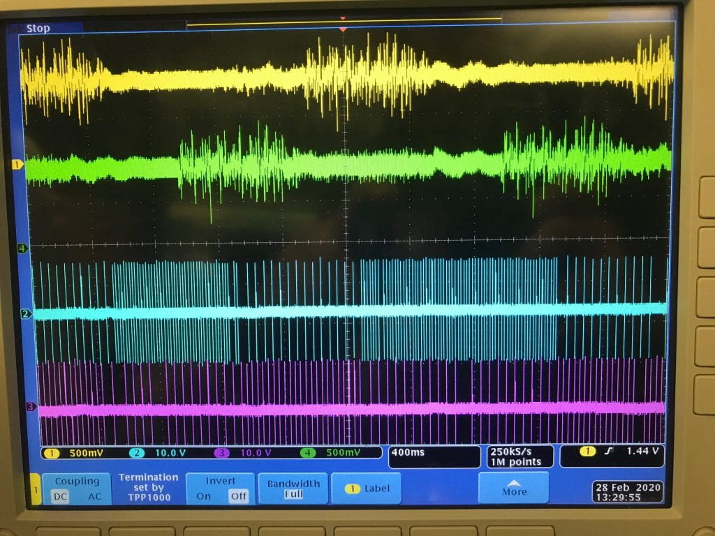
The figure displays a demonstration of our closed-loop system performing real-time, multi-channel recording and stimulation. The green and yellow traces are EMG signals fed into the recording sensors of the chip. The information is transmitted wirelessly as input to a control algorithm that detects EMG envelopes over the chip’s multiple recording channels. The decision algorithm modulates the chips stimulation parameters to 60Hz (blue trace) and 20Hz (pink trace) when a large envelope is detected in the yellow EMG signal; and 20Hz (blue trace) and 30Hz (pink trace) when a large envelope is detected in the green EMG signal.


Successful Validation of Bi-directional Communication for Our Implant SoC

Link to Demo Video

We have successfully tested and validated the bidirectional communication links (forward and reversed) for our implant SOC and system designed for the restoration of motor functions for the spinal cord injury (SCI). The stimulation commands are issued via forward link(cyan trace) while the response to the commands is digitally transmitted and received by the customized Graphic Interface Unit (GUI) via the reversed link (green trace). The command is capable of simultaneously targeting various spinal cord segments and thus the corresponding muscles at the lower limbs by different set of waveform parameters, including frequency, skew, duration, amplitude, phase, etc. (yellow and pink traces). This is a critical step toward the realization of a closed-loop system for SCI applications.

Spinal Cord Prosthesis

We are collaborating with Huntington Medical Research Institutes on intraspinal microstimulation to restore bladder and bowel movement, and sexual function after spinal trauma. The goal is to use intraspinal microstimulation to artificially trigger the reflexes of the visceral organs, after the activation of the spinal circuitry from the brain stem is lost due to spinal cord injury or disease. We are currently designing a 32-channel implantable stimulation chip for implantation in the spinal cord. In order to study the effect of stroke on the spinal circuitry, Dr. Pikov has demonstrated a “virtual stroke” method through reversible cortical inactivation. The goal is to observe the effect of “virtual stroke” on the spinal cord through neural recording, during and after the cortical inactivation. We are currently designing a 48-channel implantable neural recording chip for implantation in the spinal cord.